
Artificial intelligence has spent years hovering over medicine as a promise — useful in theory, occasionally impressive in pilot studies, but often still distant from the bedside. On Thursday, two developments in Britain and Europe suggested that distance may be narrowing.
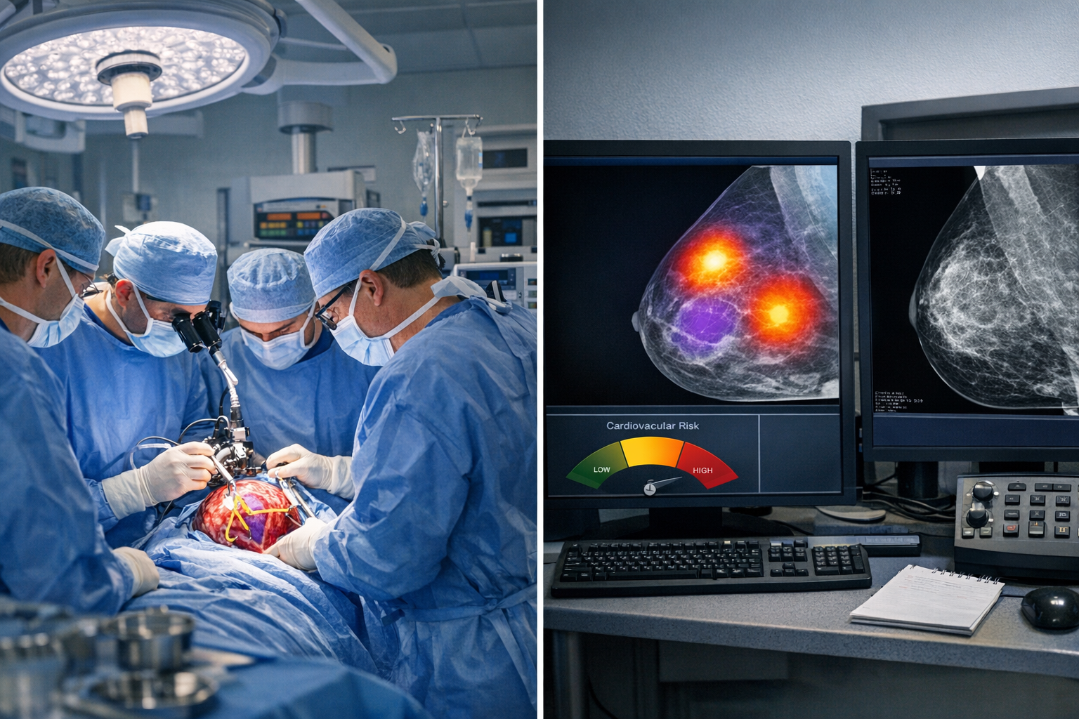
In London, surgeons said they had completed what University College London Hospitals described as the first reported real-time A.I.-assisted live brain-tumor removal, using software that analyzed the operation as it happened and flagged delicate anatomy the surgical team needed to avoid. In separate research to be presented at the European Society of Cardiology’s annual meeting, investigators reported that A.I. could mine routine mammograms for signs of cardiovascular disease, potentially turning a standard breast-cancer screening appointment into an additional warning system for women’s heart health.
The two advances are very different in setting and ambition. One unfolds in the intensity of the operating room, where a split-second decision can preserve a patient’s sight. The other could work quietly in the background of mass screening programs, drawing new clues from images already being collected. But together they point to a more tangible phase of medical A.I.: not as a replacement for clinicians, but as a tool embedded in care.
A landmark case in the operating room
The brain surgery took place in May at the National Hospital for Neurology and Neurosurgery, part of University College London Hospitals NHS Foundation Trust, but was disclosed only after the patient had recovered.
The patient, Rhys Hibbert, 48, underwent an operation to remove a pituitary tumor. According to the hospital, the A.I. system — developed at University College London and used as part of a clinical trial — analyzed the live video feed from the camera used during surgery and highlighted critical structures in real time. The goal was to help surgeons distinguish the tumor from nearby anatomy that, if damaged, could have devastating consequences.
Hospital officials said the tumor was removed and Mr. Hibbert’s sight was preserved.
That matters because pituitary surgery is among the kinds of procedures where precision is unforgiving. The gland sits deep at the base of the brain, close to the optic nerves and major blood vessels. Even for highly skilled surgeons, distinguishing tissue planes and avoiding tiny but essential structures can be difficult.
The UCL system was trained on previously annotated pituitary-surgery videos, according to the hospital, and funded by Britain’s National Institute for Health and Care Research and Google. For proponents of surgical A.I., the case offers a vivid example of what the technology could do best: serve as an extra set of eyes, constantly scanning for danger while the surgeon remains in control.
Still, a milestone is not the same as proof. The operation involved a single patient in an early-stage trial, and the central questions now are familiar ones in medical innovation: whether the system works reliably across more patients, more surgeons and more hospitals; whether it improves outcomes compared with expert surgery alone; and how regulators will assess tools that guide clinicians during live procedures.
A new use for a familiar screening test
The second development may ultimately affect far more patients, if it holds up.
Researchers reported that A.I. analysis of routine mammograms could identify women with hypertension, ischemic heart disease or a prior stroke. The findings suggest that images taken to look for breast cancer may also contain cardiovascular signals that are not routinely used in clinical practice.
That possibility is especially significant because heart disease remains the leading cause of death in women worldwide, and one that is often underdiagnosed or recognized later than it is in men. Symptoms can present differently, risk can be underestimated, and women’s cardiovascular health has historically received less targeted attention.
Using mammograms as a second screening window could change that equation. Breast screening is already deeply embedded in health systems, and millions of women undergo mammography every year. If A.I. can reliably detect vascular changes or patterns associated with cardiovascular disease from those same images, clinicians may be able to identify high-risk patients without ordering additional scans.
The new research remains preliminary. It is due to be presented at a medical conference and is retrospective, meaning it looks back at existing data rather than testing the approach prospectively in real time. Before such a tool could become standard practice, researchers would need to show that it performs consistently in broader populations, that it improves decision-making, and that it does not trigger waves of unnecessary anxiety or follow-up testing.
But the idea is not emerging from nowhere. Earlier this year, a peer-reviewed study published in the European Heart Journal examined 123,762 women and found that A.I.-quantified breast arterial calcification on mammograms added prognostic value for major adverse cardiovascular events and mortality beyond standard risk scoring. That research strengthened the case that mammograms may hold clinically useful cardiovascular information that has largely gone untapped.
From hype to clinical utility
For years, some of the most publicized claims about medical A.I. centered on what algorithms might someday diagnose better than doctors. The more immediate path now appearing in hospitals is narrower, but perhaps more consequential: decision support in moments of risk, and opportunistic screening built into care patients already receive.
In the London operating room, that meant software identifying anatomy that surgeons should not touch. In breast screening, it could mean an alert that a woman who came in to check for cancer may also need an assessment of her cardiovascular risk.
Both cases also underscore the limits of the current moment. The neurosurgery result is a landmark but early one. The mammogram research is promising but not yet practice-changing. Neither finding resolves the broader concerns that have shadowed medical A.I., including bias in training data, uneven performance across populations, liability when systems fail, and the risk that hospitals adopt expensive technology before its benefits are clear.
Yet medicine often changes not through grand declarations but through specific, credible uses that solve real problems. Preserving a man’s sight during delicate brain surgery is one such use. Finding heart risk in a test women are already having may be another.
What happens next will depend less on headlines than on evidence: larger trials, peer-reviewed validation, regulatory scrutiny and the willingness of clinicians to trust systems that must prove they can help without distracting, overwhelming or misleading. But after years in which medical A.I. was discussed mainly as an approaching transformation, Thursday’s developments offered something more concrete — a glimpse of how it might enter everyday care.
Sources
Further reading and reporting used to add context:
- https://www.theguardian.com/society/2026/aug/27/mammograms-heart-disease-in-women-study
- https://www.theguardian.com/technology/2026/aug/27/london-neurosurgeons-ai-assisted-operation-brain-tumour
- https://www.thenationalnews.com/news/uk/2026/08/27/ucl-surgeons-pioneer-ai-aided-brain-tumour-operation/
- https://www.eurekalert.org/news-releases/1141382
- https://www.nature.com/articles/s41591-026-04277-x
- https://londoncrimenews.co.uk/latest/london-surgeons-use-ai-in-world-first-brain-tumour-operation
- https://onthewire.ai/article/london-surgeons-removed-a-brain-tumour-with-real-time-ai-guidance-a-world-first
- https://saudigazette.com.sa/article/664105/life/worlds-first-patient-to-undergo-live-ai-assisted-brain-surgery-has-tumor-removed
- https://www.the-independent.com/news/health/brain-tumour-treatment-surgery-ai-rhys-hibbert-b3040113.html
- https://www.eurekalert.org/news-releases/1118661
- https://www.theintelligent.ec/health/2026/london-neurosurgeons-complete-world-first-live-ai-assisted-brain-tumour-removal
- https://www.jmir.org/2026/1/e99154/
- https://www.reddit.com/r/aiwars/comments/1w00hkb/ai_enters_the_operating_room_london_brain_surgery/
- https://www.reddit.com/r/DefendingAIArt/comments/1w00hjc/ai_enters_the_operating_room_london_brain_surgery/
- https://www.jmir.org/2026/1/e99154/PDF
- https://www.reddit.com/r/ArtificialInteligence/comments/1vzl1oy/first_patient_to_have_brain_surgery_with_realtime/
- https://www.reddit.com/r/UpliftingNews/comments/1vzxwba/ai_can_detect_heart_disease_in_women_using/
- https://www.reddit.com/r/accelerate/comments/1vznzkx/worlds_first_patient_to_undergo_live_aiassisted/
- World-first AI-assisted brain surgery saves patient’s sight
- https://esc365.escardio.org/journal/92097
- https://www.ucl.ac.uk/brain-sciences/ion/research/queen-square-clinical-trials-centre/current-trials-list-across-queen-square/neurosurgery
- https://www.escardio.org/guidelines/scientific-documents/recommended-reading/cardio-oncology/
- https://www.escardio.org/news/press/press-releases/ai-can-predict-risk-of-serious-heart-disease-from-mammograms/
- https://www.ucl.ac.uk/brain-sciences/ion/research/research-departments/department-translational-neuroscience-and-stroke/brain-tumour-group
- https://discovery.ucl.ac.uk/id/eprint/10199474/7/Marcus_Artificial%20Intelligence%E2%80%93Assisted%20Surgical%20Scene%20Recognition_VoR.pdf
- https://www.ucl.ac.uk/brain-sciences/events/2026/mar/im-brain-surgeon-ask-me-anything
- https://www.ucl.ac.uk/brain-sciences/ion/research/clinical-divisions/division-neurosurgery/clinical-trials
- https://www.ucl.ac.uk/hawkes-institute/ucl-medical-image-computing-summer-school-medicss/medicss-2023/robotics-and-ai-neurosurgery
- https://esc365.escardio.org/journal/94213
- https://discovery.ucl.ac.uk/10200059/1/s41746-024-01273-8.pdf
- https://discovery.ucl.ac.uk/id/eprint/10227805/
- https://www.ucl.ac.uk/brain-sciences/sites/brain_sciences/files/2023_annual_report_for_the_ucl_queen_square_institute_of_neurology_division_of_neurosurgery_.pdf
- https://www.ucl.ac.uk/ion/sites/ion/files/2019-2020_bi-annual_division_of_neurosurgery_report.pdf
- https://esc365.escardio.org/journal/93607
- https://www.ucl.ac.uk/brain-sciences/sites/brain_sciences/files/2025-08/2024%20Annual%20Report.pdf
- https://conferences.medicom-publishers.com/wp-content/uploads/2021/03/LR_ESC2019_MAIN_Non-Sponsored.pdf
- https://www.escardio.org/news/press/press-at-congresses/esc-congress-press-activities/
- https://esc365.escardio.org/journal/92035
- https://www.escardio.org/events/congresses/esc-congress/scientific-programme/
- https://www.escardio.org/static-file/Escardio/Congresses/ESC%20Congress/Call%20for%20science/ESC%20Congress%202026_Topic%20List%20for%20Abstract%20Submission.pdf
- https://www.escardio.org/events/congresses/esc-congress/helpful-information/our-data-science-activities-at-esc-congress/
- https://www.escardio.org/topics/digital-health-ai/
- https://www.escardio.org/news/news-room/cv-health-checks-at-esc-congress-2026/
- https://www.escardio.org/news/press/press-releases/
- https://www.escardio.org/communities/working-groups/e-cardiology/news/editorial-february-2026/
- https://esc365.escardio.org/journal/92251
- https://www.escardio.org/static-file/Escardio/Congresses/ESC%20Congress/Call%20for%20science/ESC%20Congress%202025_Topic%20List.pdf
- https://www.escardio.org/static-file/Escardio/About%20the%20ESC/Annual-Reports/esc-annual-report-2025.pdf
- https://www.escardio.org/static-file/Escardio/Congresses/ESC%20Congress/Documents/2022/ESC%20Congress%202022_Main%20Topics%20for%20Clinical%20Case%20Submission.pdf









Leave a Reply